ENDOCRINE INVESTIGATIONS
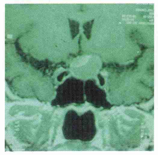 MRI is widely available at both hospitals and is routinely used in the investigation and follow-up of our
pituitary and neuro-endocrine patients. There are joint neuroradiological, neurosurgical and endocrine meetings to discuss the management of the pituitary tumours held a Charing Cross
Hospital.
MRI is widely available at both hospitals and is routinely used in the investigation and follow-up of our
pituitary and neuro-endocrine patients. There are joint neuroradiological, neurosurgical and endocrine meetings to discuss the management of the pituitary tumours held a Charing Cross
Hospital.
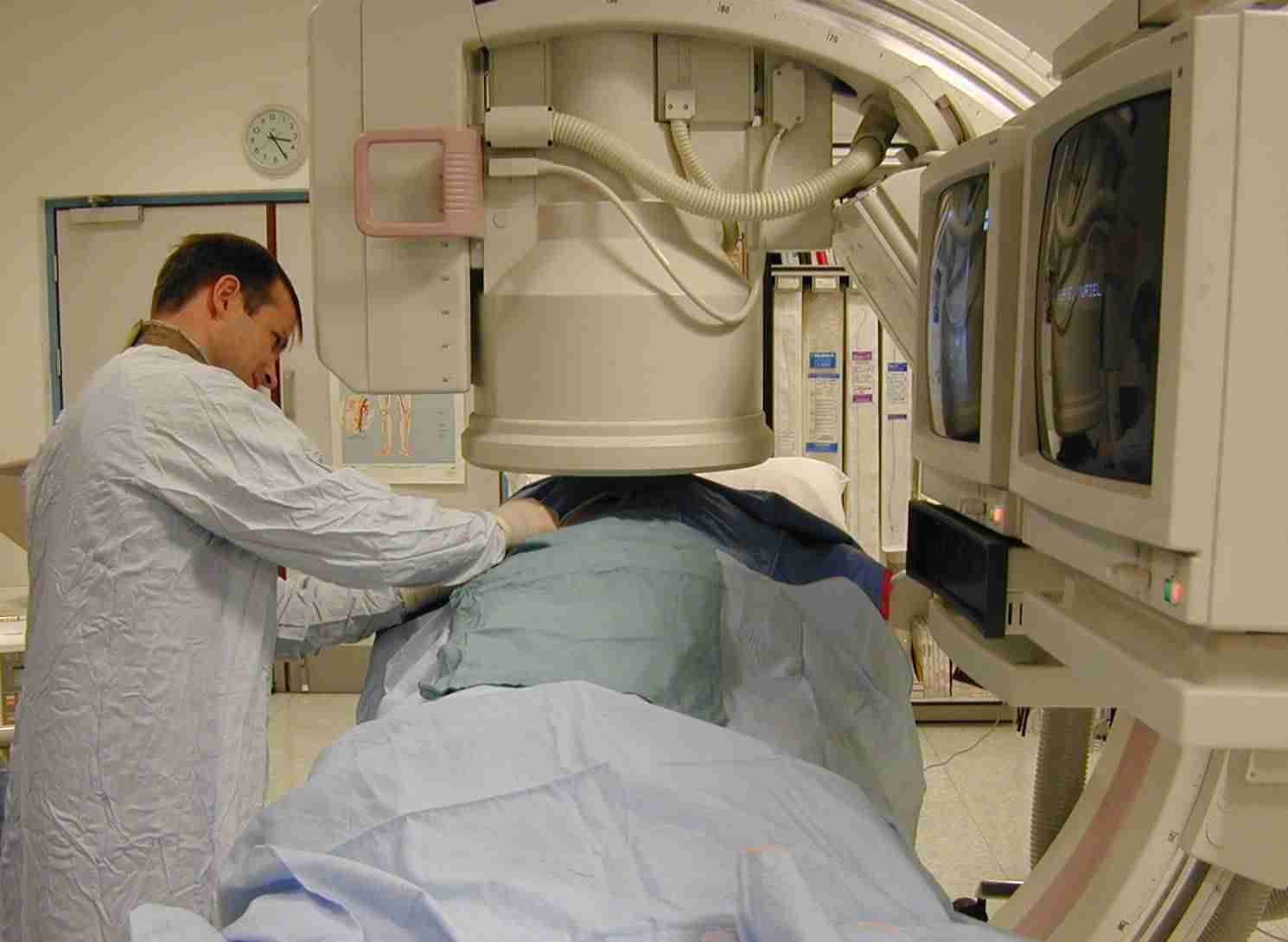 Methods for the localisation of endocrine tumours have been perfected at the Hammersmith Hospital. There is no part of the anatomical arterial or venous tree that remains inaccessible for endocrine sampling. Many sophisticated procedures are undertaken
routinely within the department of diagnostic radiology.
Methods for the localisation of endocrine tumours have been perfected at the Hammersmith Hospital. There is no part of the anatomical arterial or venous tree that remains inaccessible for endocrine sampling. Many sophisticated procedures are undertaken
routinely within the department of diagnostic radiology.Inferior Petrosal Sinus SamplingPerformed on all patients with Cushing's syndrome prior to surgery. This technique allows central and peripheral lesions to be identified. The SpRs help both with the procedure and the interpretation of the results. |
Calcium Stimulation Tests for localisation of insulinomas (page 10)
HOME